

You've lost a molar, your calendar is packed, and you want a clear answer before you book anything: can the dental implants procedure replace the tooth in one appointment, or are you committing to months of treatment? The honest answer depends less on a standard script and more on your bone, gums, bite, anatomy, and the restoration you need.
A dental implant is a replacement tooth root that supports a crown, bridge, or denture. Modern implant dentistry has become a mainstream restorative option. In a large U.S. population study of adults missing at least one tooth, implant prevalence increased from 0.7% in 1999-2000 to 5.7% in 2015-2016, with the authors reporting an average covariate-adjusted increase of 14% per year during that period, as summarized in this clinical overview of dental implant outcomes.
That popularity doesn't mean every case follows the same timeline. Some patients have enough healthy bone for placement soon after extraction. Others need infection control, gum treatment, site preservation, bone grafting, or a sinus lift before an implant can be supported safely. The procedure is predictable when the plan respects those biological limits.
Why the Dental Implants Procedure Is More Than a Single Visit
A busy Manhattan professional may ask, “I'm missing one tooth. Can we place the implant and crown this afternoon?” In selected cases, implant placement can be completed efficiently. The complete treatment pathway usually includes diagnosis, planning, possible site preparation, surgical placement, healing, and final restoration. Your anatomy determines how many of those stages can be combined safely.
The implant replaces the tooth root. A separate crown is designed and fitted after the surrounding bone has stabilized the fixture. Osseointegration describes the process in which bone bonds with the implant surface. That biological connection allows the final restoration to function as a fixed replacement instead of a removable appliance.
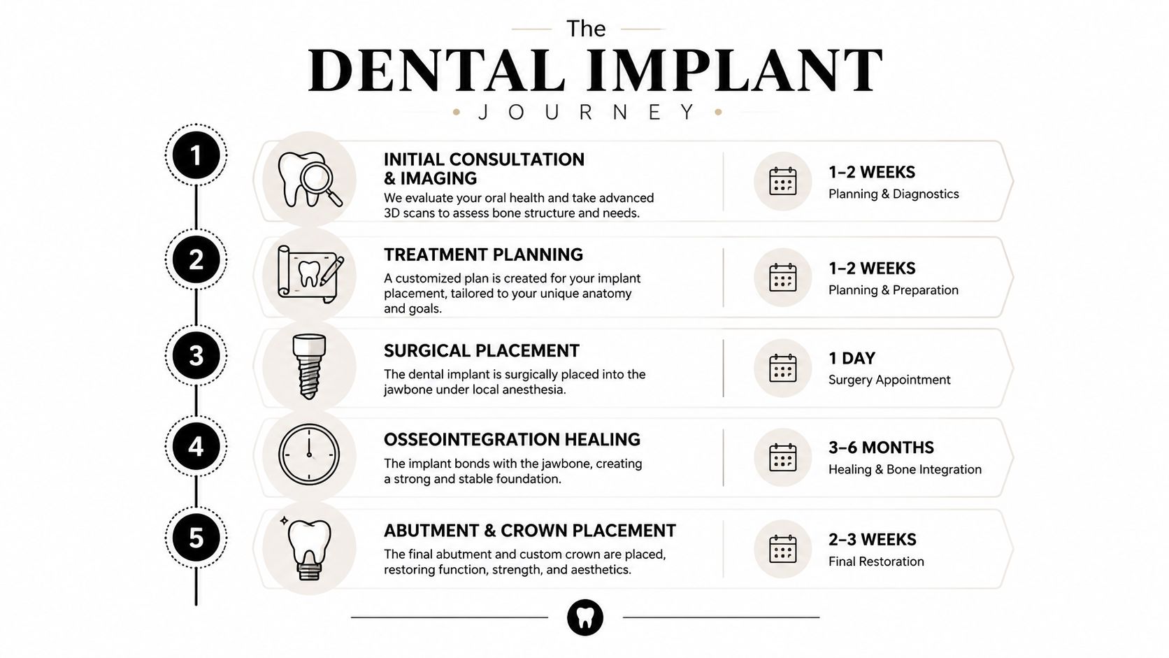
The five phases you should expect
- Consultation and imaging: The dentist assesses your oral health, bone, bite, and medical factors relevant to healing.
- Treatment planning: The team decides whether extraction, gum therapy, grafting, or a temporary tooth belongs in the sequence.
- Surgical placement: The implant is inserted into the prepared site with appropriate anesthesia.
- Healing: The implant remains protected while bone integration develops.
- Abutment and crown: The connector and final restoration are fitted, adjusted, and checked during function.
A straightforward single-tooth case may progress through these phases with few detours. A tooth missing for years can leave thinner or reduced bone. Periodontal disease may require treatment before surgery, while full-arch care requires coordination among several implants and a fixed prosthesis. These anatomical and biological differences explain why one patient may receive same-day temporary teeth while another needs staged care over several months.
Practical rule: The quickest appointment is not necessarily the quickest safe plan. Choose the shortest timeline that protects implant stability, tissue health, and a manageable bite.
Ask your dentist to provide the sequence in writing. It should state what happens if the implant needs additional healing time and whether a temporary restoration is required. For financial planning, review these factors that influence dental implant cost. The useful question is, “What must be true in my mouth before the final tooth is safe to place?”
The Consultation and 3D Imaging Phase
The first appointment should be a diagnostic visit, not a sales presentation. Your dentist needs to understand the missing tooth site, the surrounding teeth, your gums, your bite, and your medical history before recommending immediate placement or preparatory treatment.
A 3D cone-beam scan can show the anatomy that ordinary two-dimensional images may not reveal clearly. The dentist evaluates the height and width of available bone, the position of the mandibular nerve, the relationship between upper back teeth and the maxillary sinus, and the shape of the ridge. The scan also helps identify neighboring roots, hidden infection, and anatomical limitations that can change the surgical approach.
What happens during the appointment
Bring an updated medication list, information about previous dental treatment, and details about medical conditions that affect healing. Expect direct questions about:
- Smoking or nicotine use: These habits can influence healing and tissue health.
- Diabetes: Your dentist may need to understand how well it's controlled.
- Medications: Bisphosphonates and other drugs can affect surgical planning.
- Grinding or clenching: Excessive bite forces can influence the restoration design.
- Gum history: Active periodontal inflammation should be addressed before implant placement.
Your dentist may use an intraoral camera to show the extraction site, gum margins, fractured teeth, or neighboring restorations. Digital scanning can create an impression-free model for planning the crown and evaluating how the proposed tooth will meet the opposing teeth.
How to judge the diagnostic process
You should leave knowing whether you're a candidate, what preparation is needed, how many broad phases are expected, and what alternatives exist. A provider who proposes surgery without examining bone, nerve location, sinus anatomy, gum health, and bite is skipping information that can affect safety and long-term function.
A panoramic image may be useful, but it doesn't answer every three-dimensional question. You can also review how imaging supports dental diagnosis in this guide to dental X-rays and their clinical role. The scan isn't just a picture for the file. It's the map used to determine whether immediate placement, delayed placement, grafting, or a different restoration makes sense.
Surgical Placement and the Osseointegration Waiting Period
The surgical appointment is often shorter than patients expect. For a single tooth, implant placement commonly takes 30 to 90 minutes, depending on the site and the required preparation. The procedure is usually performed with local anesthesia, so patients generally feel pressure, vibration, and movement rather than sharp pain.
The dentist anesthetizes the area, accesses the bone, prepares the implant site with controlled drilling, and inserts the fixture. A healing component or temporary restoration may be used according to the stability of the implant, the location of the tooth, the condition of the gums, and the loading plan.
What healing involves
The most important part begins after you leave. Bone must integrate with the implant before the final crown receives normal chewing forces. Healing commonly takes 3 to 6 months, although the appropriate interval depends on primary stability, bone quality, the location of the implant, whether grafting was performed, and the overall treatment design.
During the early phase, swelling and tenderness can occur. Follow your dentist's instructions about cold packs, medication, oral hygiene, diet, and physical activity. Soft foods can protect the site while the tissues settle, and you shouldn't test the implant by repeatedly touching it with your tongue or applying pressure during chewing.
The implant may feel fine before integration is complete. That's why follow-up appointments matter. Your dentist may examine the tissues, assess the restoration or healing component, review your bite, and use clinical judgment or imaging before moving to the abutment and crown.
Feeling comfortable doesn't prove that the implant is ready to load. Stability must be evaluated clinically.
Long-term evidence supports judging implants by multi-year performance rather than by how the site looks immediately after surgery. A systematic review estimated 10-year implant survival at 96.4%, with a prediction interval of 91.5% to 99.4%, while a sensitivity analysis estimated 93.2% survival after accounting for patients lost to follow-up, as reported in this PubMed review of long-term implant survival.
For a visual explanation of the recovery phase, see this overview of the dental implant healing process.
When Bone Grafting and Site Preservation Change the Timeline
Bone determines whether an implant can be placed and stabilized where the tooth used to be. After a tooth has been missing, the ridge can become narrower or shorter. Periodontal disease, trauma, infection, natural anatomy, and the position of the maxillary sinus can also limit the available support.
That's why two people replacing the same tooth may receive different recommendations. One may qualify for placement after extraction because the socket walls and surrounding bone remain favorable. Another may need to rebuild the site first.
Three types of preparation
- Socket preservation: A graft is placed in the extraction socket to help maintain the ridge while it heals.
- Ridge augmentation: The dentist adds material where the ridge lacks adequate width or height for implant support.
- Sinus lift: In the upper back jaw, the sinus membrane may need to be carefully moved to create room for bone beneath it.
These procedures aren't interchangeable, and they don't belong in every treatment plan. The scan, clinical examination, infection status, and intended crown position determine whether preparation is necessary.
Immediate treatment versus staged treatment
Immediate implant placement can be appropriate when the extraction site, surrounding bone, gum tissue, and infection status are favorable. Immediate loading, meaning the use of a provisional or final restoration soon after placement, requires dependable stability and careful control of chewing forces. It isn't a shortcut that can be applied safely to every patient.
A straightforward single-tooth plan may be completed in 4 to 6 months, while complex full-arch rehabilitation may require 12 to 18 months of staged procedures, depending on preparation and healing needs. Those timeline figures are treatment scenarios, not guarantees, and the clinical literature emphasizes that poor primary stability, inadequate bone quality, immediate post-extraction placement, and atrophic jaws can alter outcomes, as discussed in this review of implant complications and risk factors.
The trade-off is clear. Staging adds visits and waiting, but it can create a more reliable foundation. If your dentist recommends grafting, ask to see the scan and the specific defect being treated. If another provider says grafting is unnecessary, ask how that decision affects implant position, stability, esthetics, and the long-term maintenance plan. You can also discuss gum anatomy and recession with a dentist familiar with gum recession treatment.
Understanding Real Complication Rates and Risk Factors
Implants have strong long-term outcomes, but “high success” doesn't mean “no complications.” The relevant risk depends on whether the problem occurs before loading, after the implant has been functioning, around the tissues, or in the crown and supporting components.
A systematic review found pre-loading implant loss of about 2.5%. For fixed reconstructions, implant loss during function was reported at about 2% to 3%, while overdenture therapy could exceed 5% loss over 5 years, according to this systematic review of implant complications. The same evidence base reported an overall complication frequency of 13.9% in one large retrospective cohort, including inflammatory complications at 10.2% and prosthetic complications at 2.7%.
More recent clinical data from 150 implant surgery cases reported infection in 15.3%, peri-implantitis in 12.0%, implant failure in 8.0%, and no complications in about 66.7% of cases, as reported in this clinical analysis of implant surgery complications. These findings shouldn't be treated as a universal prediction because patient selection, procedures, follow-up, and definitions vary.
Risks that deserve a candid discussion
Possible complications include infection, inflammation around the implant, incomplete integration, gum recession, persistent sensitivity, crown or abutment loosening, bite discomfort, bleeding, wound separation, nerve injury, and sinus complications. The risk profile changes with smoking, uncontrolled systemic disease, poor plaque control, untreated periodontal disease, inadequate bone, and heavy clenching.
Some risks are managed before surgery. Your dentist may stabilize gum disease, review medications, adjust the bite plan, or recommend a bridge or partial denture instead. An implant isn't automatically the right choice just because a tooth is missing.
Warning signs after surgery
Mild soreness and swelling can occur during early healing. Contact your dentist promptly if pain worsens instead of improving, swelling continues to increase, bleeding doesn't settle, discharge develops, fever appears, or the implant feels mobile. A sudden bite change also deserves review because excessive force can affect a healing site or restoration.
The useful question isn't “What's your success rate?” Ask, “What are the main risks in my anatomy, how will you reduce them, and what will you do if healing doesn't follow the expected course?”
Final Restoration and Long-Term Maintenance Protocols
Once integration is confirmed and the gum tissue has healed sufficiently, the restorative phase begins. The abutment connects the implant to the crown, bridge, or denture. Your dentist records the implant site, adjacent teeth, opposing bite, and appearance required for the final restoration.
Digital scanning can produce a detailed model without conventional impression material. The laboratory uses that record to design the restoration, while your dentist evaluates contact points, bite, emergence profile, and access for cleaning. A follow-up adjustment may be needed after you start chewing. A crown can appear correct yet feel high or uneven under function.
The implant has no periodontal ligament and provides different sensory feedback from a natural tooth. Inflammation around it may therefore develop without the same warning sensations you would associate with a natural tooth.
A practical maintenance routine
- Clean daily: Brush around the crown and gumline. Use an interdental brush or water flosser if your dentist recommends it.
- Monitor the tissue: Bleeding, swelling, tenderness, pus, persistent bad taste, or a changed gum contour warrants an examination for inflammation.
- Protect the bite: If you grind or clench, ask whether a custom night guard fits your restoration and bite.
- Keep professional visits: Implant surfaces and prosthetic contours may require instruments and techniques designed for implant maintenance.
- Report movement: A loose crown and a mobile implant represent different problems. Both require examination, not home adjustment.
Long-term evidence supports regular follow-up, while also showing that survival is not identical for every implant or patient. In a 10,871-implant clinical study, implant-level survival was 98.9% at 3 years, 98.5% at 5 years, 96.8% at 10 years, and 94.0% at 15 years. Patient-level survival was 97.4% at 3 years and 92.5% at 10 years, as summarized in this report on long-term dental implant survival.
These figures describe implant survival, not a guarantee that the crown will never need repair or replacement. Before treatment, confirm who provides maintenance, what the warranty covers, how prosthetic repairs are handled, and whether follow-up remains available if you move or change insurance. For home care, review these practical tips for keeping dental implants clean.
Choosing the Right Provider and Timing Your Treatment
A good implant provider should make the treatment variable for the right reasons, not vague for avoidable ones. Look for a practice that can evaluate bone and soft tissue, use appropriate imaging, explain restorative design, and coordinate surgery with the final crown or prosthesis.
Ask these questions before accepting a plan:
- Who performs each phase? Clarify whether diagnosis, surgery, restoration, and maintenance are handled by one team or divided among referrals.
- What does the scan show? Ask the dentist to identify bone width, nerve location, sinus anatomy, and the proposed implant position.
- Why is the timeline this long? A credible plan should explain healing, grafting, infection control, or staged full-arch work in clinical terms.
- What happens if the implant doesn't integrate? Understand the response, additional treatment, and financial responsibility before surgery.
- How will the final crown be maintained? Ask about access for cleaning, bite adjustments, repairs, and replacement of prosthetic components.
How to compare treatment plans
| Treatment plan feature | Appropriate reason for variation | Question to ask |
|---|---|---|
| Advanced imaging | More detailed assessment of bone and anatomy | “What decision will this scan help you make?” |
| Bone grafting or site preservation | Insufficient ridge support or tissue management | “What specific defect are you correcting?” |
| Different implant or crown components | Restorative design, esthetics, or bite demands | “Why is this component suited to my case?” |
| Longer timeline | Healing, infection control, or staged rehabilitation | “Which biological step prevents earlier loading?” |
| Lower quoted fee | A different scope, material, or follow-up arrangement | “What is included, and what is excluded?” |
Busy professionals often try to compress appointments around travel, presentations, and work deadlines. That can work for consultations, scans, and some restorative visits, but it shouldn't dictate biological healing. Teledentistry may help with an initial discussion or follow-up questions, while surgical candidacy still requires an in-person examination and appropriate imaging.
Paul L. Gregory, DDS provides implant dentistry, bone grafting, site preservation, All-on-4 full-arch treatment, digital scanning, intraoral imaging, and related restorative care at its Midtown Manhattan practice. That makes it one option for patients who want implant planning and broader dental care coordinated in one office.
Start by scheduling a diagnostic consultation rather than shopping for a same-day promise. Bring your medical history, ask for a written sequence of visits, and get a second opinion if the provider can't explain why your anatomy requires immediate placement, grafting, delayed loading, or an alternative such as a bridge or partial denture.
If you're considering dental implants in Manhattan, visit Paul L. Gregory, DDS for an evaluation that connects 3D diagnostics, surgical planning, restoration, and long-term maintenance. The team can review whether you need site preservation, bone grafting, single-tooth replacement, or a full-arch option, then help you plan treatment around your schedule.