

You notice it while brushing your teeth. One tooth looks a little longer than the others, cold water sends a sharp sensation through the root, or a faint pink line appears in the sink. Sometimes the change is so gradual that you only recognize it after seeing an older photograph of your smile.
That moment doesn't automatically mean you need surgery. Gum recession treatment is a matching problem, not a one-size-fits-all purchase. The right response depends on why the tissue moved, how much attachment remains, whether the exposed root has a cervical lesion or filling, and whether the area is stable after careful cleaning and habit changes.
Why Gum Recession Matters More Than You Think
A gumline can change without warning. One morning, a tooth may look longer, cold water may cause a sharp sensation, or brushing may leave a small trace of blood. The visible change is only part of the picture. As the gum margin moves toward the root, that newly exposed surface can react to temperature, touch, brushing, and acidic foods.
The crown is protected by hard enamel. Much of the root is covered by cementum, a thinner surface that is less resistant to wear and irritation. That difference explains why a small-looking recession may create sensitivity or make brushing uncomfortable. It does not, by itself, mean surgery is needed.
Recession is common and does not occur only in people who neglect oral hygiene. A 2022 systematic review and meta-analysis included 15 population-based studies and 37,460 participants. It estimated pooled prevalence at 78.16% using the minimal reported threshold and 84.92% at a threshold of at least 1 millimeter, with buccal recession present in 75.42% of participants. These findings appear in the updated review of gingival recession prevalence and treatment.
Earlier U.S. data cited in an NIH review estimated that 23% of adults aged 30 to 90 had one or more tooth surfaces with at least 3 millimeters of recession. Another survey found recession at one or more sites in more than half of the population. Recession also becomes more common with age, so a changing gumline deserves assessment rather than dismissal.
What early evaluation can clarify
A dentist or periodontist can separate abrasion, inflammation, periodontal attachment loss, clenching, tooth position, and combined causes. The examination can also identify root decay, a non-carious cervical lesion, a defective filling, changes in bone support, and the thickness of nearby tissue.
That information makes treatment a matching decision. Stable, mild recession may need gentler brushing, plaque control, sensitivity protection, and observation. Active damage, an exposed lesion, or inadequate tissue support may call for grafting, restorative care, or both. A professional dental cleaning and exam establishes a useful baseline and reduces guesswork.
A calm first step: Find out whether the tissue is still being damaged, or whether the recession is stable and needs protection and monitoring.
How Gums Recede and How Dentists Classify the Change
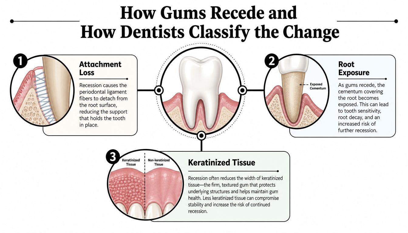
Healthy gum tissue fits like a snug collar around each tooth. It meets the enamel, seals the margin, and works with the periodontal attachment beneath it to protect supporting structures. As the gum edge shifts toward the root, the tooth looks longer and the newly exposed surface becomes more vulnerable to irritation.
Three changes help explain what has happened:
- Attachment loss means the connection among the gum, tooth, and surrounding periodontal structures has moved or been damaged.
- Root exposure means cementum or another root surface is open to the mouth, toothbrush contact, temperature changes, and plaque.
- Keratinized tissue changes involve the amount and quality of the firmer gum tissue around the tooth.
Why classification affects the plan
Classification gives the dentist a practical forecast. It helps estimate how much root coverage may be realistic and whether the nearby tissue can be moved over the exposed root predictably. The assessment includes recession depth, attachment between the teeth, supporting tissue, tooth position, and local gum biotype.
Older systems commonly used Miller classifications. Newer periodontal systems include RT1, RT2, and RT3 categories. In plain language, recession with intact tissue and bone between the teeth usually offers a better coverage opportunity than recession accompanied by interproximal attachment loss. A broad, thick gum margin may also respond differently from a narrow, thin one.
A concrete example makes the distinction easier. A patient with a thick biotype and a Miller Class I recession on an upper canine may have a favorable chance of complete root coverage. A patient with thin tissue and bone loss between the teeth has a different prognosis, even if the exposed root looks similar in a mirror. The right treatment therefore depends on the recession type and the tooth's condition, not on a one-size-fits-all graft promise.
The measurements behind the diagnosis
During an evaluation, the clinician may record recession depth, probing depth, clinical attachment level, tissue thickness, keratinized tissue width, plaque levels, and bleeding. Photographs and digital imaging can help show whether the gumline is changing over time.
The key question is not just whether the gum can be covered. It is whether the cause is still active, whether the root has decay or a non-carious cervical lesion, and whether the surrounding tissue can support a stable result. Mild, stable recession may need protection and monitoring. A damaged root surface, inadequate tissue, or ongoing attachment loss may require grafting, restorative treatment, or both.
What Causes Gums to Pull Back Over Time
Recession usually reflects more than one influence. One patient may brush too forcefully over naturally thin tissue. Another may have inflammation around a defective restoration. A third may clench heavily while a tooth sits outside the protective contour of the dental arch.
Mechanical irritation
A hard-bristled brush, excessive pressure, or a back-and-forth sawing motion can repeatedly traumatize the gum margin. The same problem can occur when someone focuses intensely on a sensitive tooth and scrubs it harder, hoping to make it cleaner. The tissue needs plaque removal, but it doesn't benefit from force.
A dentist may recommend a soft-bristled manual brush or an electric brush with a pressure sensor, along with small, controlled movements. The correction is behavioral, not merely a new product.
Thin tissue and tooth position
Some people naturally have a thin gum biotype, a prominent tooth root, a shallow vestibule, or limited protective tissue. Orthodontic movement can also place a tooth in a position where the outer bone and gum covering are less substantial. These anatomical features don't mean recession is inevitable. They do mean the clinician must assess the tissue before moving teeth or planning cosmetic work.
Inflammation and periodontal disease
Plaque and calculus can inflame the gum and damage the attachment and bone that support a tooth. In that setting, grafting alone doesn't address the active disease. The patient first needs inflammation control, professional debridement, home-care coaching, and a maintenance plan.
Force from clenching
Clenching and grinding can overload teeth and the tissues around them. Force may not be the original cause, but it can make a vulnerable site harder to stabilize. A clinician may evaluate bite contacts and discuss a custom night guard when nighttime clenching is part of the pattern.
Restorative, orthodontic, and lifestyle factors
Overhanging fillings can trap plaque near the gumline. Cervical restorations may alter the contour that a graft must blend with. Tobacco use can impair the local environment and make periodontal control more difficult. These factors need to be addressed as part of treatment, not treated as side notes.
Treating the cause is part of treating the recession. A graft placed over a site that continues to face plaque, pressure, abrasion, or an unstable restoration is being asked to work against the same forces that created the problem.
Non Surgical Gum Recession Treatment You Start With Today
Non-surgical care is the starting point for nearly every recession evaluation, even when surgery may eventually be appropriate. It reduces inflammation, lowers sensitivity, removes local irritants, and reveals whether the tissue is stable enough to observe or repair.
Build a gentler cleaning routine
Use a soft-bristled brush and light pressure. Aim the bristles toward the gumline, but don't scrub the margin as if you're polishing a stain from a countertop. Small circular or short vibrating motions are usually more appropriate than aggressive horizontal strokes.
Clean between the teeth every day with floss or an interdental brush sized to fit comfortably. Mouthwash can support a routine, but it doesn't replace mechanical plaque removal. If a clinician recommends an antimicrobial rinse, ask how long to use it and whether it may stain teeth or alter taste.
Control sensitivity without hiding the cause
Desensitizing toothpaste can reduce symptoms over time. Fluoride varnish or another professionally selected fluoride treatment may help strengthen vulnerable root surfaces. These measures can make daily life easier, but they won't physically move the gum back over the root.
Acid exposure also matters. Frequent acidic drinks and foods can soften exposed root surfaces, especially when someone brushes immediately afterward. Your dentist can help separate sensitivity caused by exposure from sensitivity caused by decay, a crack, or an inflamed pulp.
Remove plaque traps
If calculus, an overhanging restoration, or a rough root surface keeps irritating the area, home care may not be enough. Scaling and root planing can remove deposits below the gumline and smooth surfaces that collect plaque. Learn more about periodontal scaling and root planing before assuming that every exposed root needs a graft.
Non-surgical treatment can stop active inflammation and improve comfort. It generally can't recreate lost gum tissue or close a deep defect by itself. Surgery becomes more reasonable when recession progresses, sensitivity remains difficult to control, the root is at risk, the patient wants predictable cosmetic coverage, or the tissue needs reinforcement around a restoration or orthodontic plan.
Surgical Options That Restore Coverage and Comfort
Recession surgery is a matching problem, not a one-size-fits-all graft purchase. The defect's shape, the amount and thickness of remaining gum, the position of the tooth, and the condition of its root surface all influence the plan. For clinically significant recession, predictable treatment often combines a flap design with a subepithelial connective tissue graft. The graft supplies additional tissue beneath or beside the existing gum, while the flap or tunnel places that tissue over the exposed root.
Two established approaches
A coronally advanced flap, often combined with connective tissue grafting, moves the gum toward the crown of the tooth. It may fit one recession defect or several neighboring defects when the tissue can move without excessive tension. The tooth must also have anatomy that allows the gum to rest securely in its new position.
A tunneling approach forms a passage beneath the gum, allowing a graft to slide into place while preserving the outer tissue. It can be useful for multiple adjacent recessions and may avoid some vertical incisions. Surgical access, tissue thickness, root position, and the clinician's ability to stabilize the graft still determine whether it is appropriate.
A contemporary review of periodontal plastic surgery procedures identifies coronally advanced flap and tunneling approaches combined with connective tissue grafting as reliable options for single and multiple recession defects. Evidence syntheses also associate root-coverage surgery with improvements in recession depth, clinical attachment level, and keratinized tissue width.
How the approaches compare
| Technique | What the evidence suggests | Where it may fit |
|---|---|---|
| Connective tissue grafts | Mean root coverage of 94% to 98% and complete coverage of 68% to 90% across studies | Defects where predictable coverage and greater tissue volume matter |
| Coronally advanced flap with graft versus flap alone | Greater recession reduction and more complete coverage when grafting is added | Suitable defects with adequate tissue and favorable anatomy |
| Acellular dermal matrix | Often comparable with connective tissue grafting, although results vary by defect and study | Patients who prefer to avoid a palatal donor site |
| Hyaluronic acid as an adjunct | Slight benefits in some parameters, without an overall significant advantage over connective tissue grafting | Selected cases where the clinician believes an adjunct may help |
The root-coverage figures come from a systematic review of root-coverage procedures. It also found that a coronally advanced flap with connective tissue graft achieved complete coverage more often and reduced recession more than several alternatives, including flap alone, guided tissue regeneration, and biomaterial-based approaches.
A 2025 meta-analysis found only slight benefits from hyaluronic acid in some parameters, with no significant overall advantage over connective tissue grafting. A separate 2025 systematic review reported that acellular dermal matrix was often as effective as connective tissue grafting, although some studies still favored the graft. The practical lesson is simple: a newer material is not automatically more predictable, as discussed in this review of hyaluronic acid and graft alternatives.
A surgical consultation should address donor-site needs, healing, tissue thickness, the desired cosmetic result, and whether complete coverage is realistic for that particular defect. A graft can add protection and comfort, but the exposed root, tooth position, and any restorative problem may require coordinated treatment rather than surgery alone.
Here is a visual overview of how root-coverage surgery is planned and performed:
Patients comparing procedures can review surgical and non-surgical periodontal treatments with their dental team before choosing a path.
Special Situations With Fillings Lesions and Orthodontics
A tooth with recession isn't always a simple exposed-root problem. The root may also have a non-carious cervical lesion, which is a worn, abraded, or chemically softened area near the gumline. It may already contain a composite filling or another cervical restoration.
That changes the treatment target. A graft can cover soft tissue, but the restoration may have an irregular edge, an overcontoured profile, a color mismatch, or a surface that prevents the gum from adapting naturally. The dentist may need to reshape or replace the restoration, treat the root surface, and coordinate the surgical position so the final tooth has a smooth emergence profile.
A 2025 systematic review found that combining periodontal surgery with restorative treatment can produce stable outcomes and a good emergence profile. It also noted that connective tissue graft root coverage can be successful on teeth with cervical fillings, as reported in the systematic review of periodontal and restorative treatment combinations.
Questions for a coordinated plan
Ask whether the filling should be adjusted before surgery, whether the lesion is active, and which surface will be visible after coverage. A plan that treats only the gum may leave the tooth sensitive or cosmetically uneven. A plan that restores only the tooth may leave thin tissue exposed to continuing irritation.
Orthodontics creates a similar planning issue. A tooth may need repositioning to improve the gum environment, or orthodontic movement may need to wait until a thin area is reinforced. A 2026 university thesis review found mild-to-moderate improvement in some recession cases during orthodontic treatment, but the evidence was limited and inconsistent, with no clear difference between fixed appliances and clear aligners, as described in the review of orthodontics and gingival recession.
If you're considering aligners, discuss gum thickness, tooth position, planned movements, and monitoring before treatment begins. A consultation about Invisalign clear aligners should include periodontal assessment when recession is already visible.
Protecting Your Results and Preventing Future Recession
A stable gumline depends on what happens after the initial treatment decision. Whether you receive desensitizing care, periodontal cleaning, restorative correction, orthodontic coordination, or root-coverage surgery, the daily routine must match the tissue's vulnerability.
A practical maintenance checklist
- Clean without trauma. Use a soft brush, light pressure, and a technique your hygienist has watched rather than described.
- Reach between teeth. Floss or use an appropriately sized interdental brush to reduce plaque where a toothbrush can't reach.
- Protect against clenching. If you wake with jaw fatigue, tooth soreness, or evidence of wear, ask whether a custom night guard is appropriate.
- Watch for changes. New sensitivity, bleeding, root visibility, a rough filling edge, or food trapping deserves attention.
- Keep professional visits. The right interval depends on inflammation, periodontal history, risk factors, and how well the area stays clean. Some patients need periodontal maintenance more often than routine preventive visits.
You can review dental and periodontal maintenance with your care team and use it as a framework for preserving stability. Tobacco avoidance and sensible management of acidic foods and drinks also reduce unnecessary stress on exposed root surfaces.
What to bring to your next appointment
Bring a short symptom history. Note whether sensitivity occurs with cold, brushing, sweets, or touch, and whether it fades quickly or lingers. Tell the clinician about clenching, tobacco use, recent orthodontics, whitening products, fillings near the gumline, and any change in brushing technique.
Ask three direct questions:
- Is the recession stable? You need to know whether monitoring is reasonable or whether progression is visible.
- What caused it? Treatment is more durable when the source of irritation is controlled.
- What result is realistic? Complete coverage, partial coverage, symptom control, and tissue thickening are different goals.
The best gum recession treatment isn't automatically the newest material or the most involved procedure. It is the plan that matches the recession classification, tissue quality, tooth condition, cause, and long-term maintenance you can sustain.
Paul L. Gregory, DDS offers examinations, digital diagnostics, periodontal cleanings, scaling and root planing, restorative care, orthodontic coordination, and maintenance planning for patients concerned about gum recession. Visit Paul L. Gregory, DDS to request an evaluation in Midtown Manhattan and discuss the right next step for your teeth and gums.