


You bite into something cold and feel a quick, sharp twinge. It disappears, but now your tongue keeps returning to that tooth, checking for a rough spot or a tiny hole. If your dentist has recommended a filling, you're probably wondering what happens after you sit in the chair, how much you'll feel, and whether the tooth will look and function normally afterward.
A modern filling is more than just plugging a hole. The dentist removes damaged tooth structure, protects the healthy portion, selects a material for the tooth's location and workload, then rebuilds the shape so it fits your bite. The American Dental Association describes direct restorative materials as being placed directly onto the tooth, including amalgam, resin-based composite, glass ionomer, and resin-modified glass ionomer (American Dental Association materials guide).
What Happens Before the Drill Comes Out
The appointment starts with questions, not drilling. Your dentist asks whether the tooth reacts to cold, sweets, pressure, or chewing, and whether the sensation fades quickly or lingers. That detail helps distinguish a small irritated area from decay that may be affecting the pulp, the living tissue inside the tooth.
The examination usually combines a mirror, a light, and a carefully used explorer. Your dentist looks for softened enamel, a visible opening, discoloration, or a rough area that catches the instrument. A bitewing X-ray can reveal decay between teeth, while a periapical X-ray shows the tooth and the bone around its root. A digital dental X-ray may also help your dentist explain the finding to you before treatment, as described in this guide to digital X-rays for teeth.
What the dentist is trying to determine
A dark mark isn't automatically a cavity that needs a filling. The dentist considers whether the lesion is active, whether the surface has collapsed, and whether the decay has moved beyond enamel into dentin. The depth and location matter because decay near the pulp requires a different level of caution than a shallow area on the outer enamel.
Your dentist may also use light transmission through the tooth, called transillumination, or a laser-based caries detection device such as DIAGNOdent. These tools support the clinical examination, but they don't replace judgment. Gentle probing, tapping the tooth, and asking you to bite can help identify a crack, inflammation around the root, or a problem that isn't explained by decay alone.
Practical rule: Before treatment begins, you should know which tooth is being treated, why it needs attention, which material is being considered, and what sensations to expect.
A typical filling visit is often planned as a focused appointment, though the actual time depends on the tooth, the number of surfaces involved, and whether the decay is deep. The dentist normally confirms the treatment plan and anesthesia approach before any instrument touches the tooth. That conversation is the right moment to mention dental anxiety, a previous difficult injection, sensitivity, or concern about the appearance of the finished restoration.
Numbing the Tooth and Removing the Decay
The first physical step is usually a topical anesthetic gel. It tastes medicinal and briefly makes the gum feel thick or numb. After that, the dentist places local anesthetic with a small injection. You may feel pressure and a short sting, but the topical gel and slow injection technique can make the moment easier.
Your lip, cheek, or tongue gradually feels heavy. The dentist waits for the anesthetic to work and tests the surrounding soft tissue before starting. If you can still feel a sharp sensation, say so. Pressure, vibration, and movement are expected during treatment. Sharp pain isn't something you should tolerate.
Keeping the tooth dry
Moisture control is central to a predictable filling. A rubber dam may surround the tooth with a thin sheet, or the dentist may use cotton rolls, cheek retractors, and suction. You'll notice the suction tube, the stretched cheek, and the sound of water collecting near the back of your mouth. These arrangements keep saliva away from the prepared surface and help protect your airway.
The high-speed handpiece produces the familiar whine and sprays water to control heat. At first, the dentist uses a diamond or carbide bur to create access through enamel. The sensation is usually vibration and pressure rather than pain. You may feel the dentist leaning against the tooth, but the anesthetic should prevent the cutting sensation.
Once the dentist reaches the decayed area, a slower handpiece and a round bur can remove soft, infected dentin. The dentist may also use caries-detecting dye to highlight tissue that needs closer evaluation. Modern selective caries removal aims to remove infected tissue while preserving firm tooth structure, particularly when decay is close to the pulp.
The bur sequence can sound intense, but the sound doesn't tell you how much tooth is being removed. Your dentist is judging the texture and color of the dentin, stopping when the remaining surface is sufficiently firm rather than removing healthy structure to create a larger cavity.
If the cavity is deep or the tooth has symptoms suggesting nerve involvement, the plan may change. A filling isn't the right treatment for every painful tooth, and a dentist who is concerned about the pulp may discuss additional testing or whether a general dentist can perform root canal treatment before proceeding.
Choosing the Right Filling Material
The material decision depends on the prepared tooth and several clinical factors. Your dentist considers the cavity's size, the tooth's position, the force of your bite, how well the area can be kept dry, your cosmetic preference, and habits such as grinding or clenching. A small cavity between front teeth places different demands on a filling than a broad restoration on a back molar.
The ADA places direct restorative materials into four categories, each with different behavior in the mouth (American Dental Association overview of direct restorations). The choice involves bonding, strength, moisture control, wear, appearance, and how much remaining tooth needs support. A dentist may also adjust the plan after examining the prepared walls and checking how the tooth meets its opposing tooth.
Composite resin
Tooth-colored composite is placed directly inside the cavity and bonded to enamel and dentin. It can blend closely with the surrounding tooth, which makes it useful when the restoration will show during speaking or smiling. Composite is commonly used for small-to-mid-size restorations exposed to moderate chewing pressure.
During placement, you may notice the dentist repeatedly drying the tooth, applying liquid materials, and using a blue curing light. The tooth must remain clean and dry while the surface is conditioned, the bonding materials are applied, and the composite is placed in increments. Saliva or blood entering the bonding field can weaken the seal, so the dentist may pause, replace isolation materials, or adjust the sequence if moisture appears.
Amalgam
Amalgam is a metal restoration with a long history in dentistry. The ADA notes that amalgam has been used for over 150 years, and an ADA patient education source says that more than 100 million Americans have received amalgam fillings. Amalgam can be durable and practical for some posterior, load-bearing situations.
It does not bond to tooth structure in the same way composite does. Its metallic appearance also makes it less appealing when the restoration is visible. The dentist may discuss how much tooth structure remains and whether the cavity shape provides enough support for this type of material.
Glass ionomer and indirect restorations
Glass ionomer can release fluoride and may suit surfaces that receive less chewing force, some pediatric situations, or use as a base or temporary restorative material. It generally does not offer the same wear resistance as composite or amalgam in a heavily loaded area.
A larger reconstruction may call for an indirect option, such as a porcelain inlay or onlay. A laboratory or digital workflow creates the piece outside the mouth before it is cemented onto the tooth. That approach can require an additional visit, but it may provide better support when a direct filling would leave the remaining cusps vulnerable.
| Material | Best for | Longevity | Key trade-off |
|---|---|---|---|
| Composite resin | Visible teeth and small-to-mid-size restorations | Technique and load dependent | Tooth-colored, but sensitive to moisture control |
| Amalgam | Some posterior, load-bearing restorations | Long clinical history | Durable and practical, but not tooth-colored and not bonded |
| Glass ionomer | Lower-load areas, selected pediatric uses, or bases | Wears faster in heavy load | Fluoride release, but less resistant to wear |
| Porcelain inlay or onlay | Larger indirect restorations | Depends on design, bite, and care | Strong esthetics and indirect fabrication, often requiring another visit |
Cost and insurance coverage can influence the recommendation, alongside the tooth's condition and the expected chewing forces. Ask how much healthy tooth remains, whether the material can be placed in a dry field, and what the dentist would choose if appearance were not a concern. For the financial side before treatment, review this guide to how much a filling costs, then request an estimate specific to your tooth.
Bonding, Layering, and Shaping the Filling
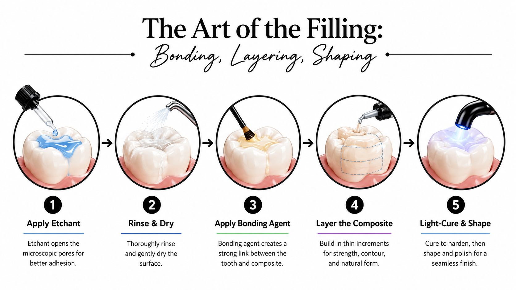
With the decay removed, you may notice the tooth feels clean and prepared while the dentist sets up the bonding steps. The enamel and dentin are prepared so the composite can adhere directly to the tooth. Depending on the bonding system, the surface may be etched and rinsed, or treated with a self-etch approach.
Etching creates microscopic irregularities, especially in enamel. The bonding agent flows into those spaces and forms the connection between tooth and composite, much like a thin primer helping two surfaces hold together. Because this layer is delicate and technique-sensitive, the dentist keeps saliva away and cures the adhesive with a blue-wavelength LED light.
Why composite goes in increments
A deep cavity is generally filled with several thin increments of composite. The dentist places one layer, directs the curing light at it, and repeats the process. Thin layers allow light to reach more of the material and help control the stress created as composite hardens and shrinks slightly.
The restoration can also follow the tooth's internal structure. A deeper layer replaces dentin, while a more translucent outer layer helps reproduce the appearance of enamel. For a small cavity, these steps may take only a short time. A broader restoration requires careful rebuilding of walls, ridges, grooves, and the contact point between neighboring teeth.
You may feel pressure from the instruments, but the filling material itself should not feel hot or sharp. Tell the dentist if your cheek, tongue, or tooth becomes uncomfortable while the layers are being placed.
The final few minutes are functional
After the composite cures, finishing burs and polishing discs refine its contours and smooth the surface. Dental floss checks whether the contact between teeth is open enough for cleaning while still limiting food impaction. Articulating paper leaves a colored mark where the opposing tooth touches.
You may be asked to tap your teeth together or move your jaw from side to side. If the filling is slightly too high, that tooth can feel as though it hits first. The dentist removes a small amount, checks the bite again, and polishes the adjusted area.
Technique affects how the restoration performs over time. A longitudinal study of posterior composite restorations reported a 5-year survival rate around 87%, an annual failure rate of 2.8%, and survival of around 82.2% at 10 years (longitudinal study of posterior composite restorations). Secondary caries, fracture, and loss of the restoration were common failure patterns. Careful isolation, incremental placement, curing, finishing, and bite adjustment therefore affect more than appearance.
When a Cavity Might Not Need a Filling at All
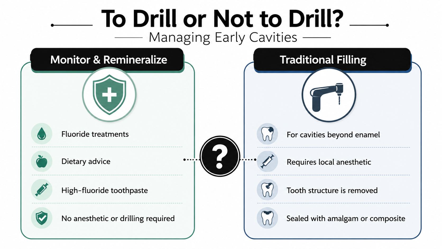
A detected lesion doesn't always mean the drill must come out that day. An early area of enamel demineralization may still have an intact surface and may be managed with fluoride, improved plaque control, dietary changes, and structured monitoring. The dentist looks for evidence that the lesion is active or progressing rather than treating every stain as a hole.
The distinction depends on depth, location, radiographic appearance, surface texture, and your personal risk of developing more decay. A shallow lesion between teeth may be approached differently from a cavitated area that traps food or has softened dentin underneath.
Conservative choices
For some early proximal lesions, resin infiltration can penetrate the porous enamel and slow or arrest progression without a conventional cavity preparation. Monitoring can also be reasonable when a lesion appears stable and the patient can return for reassessment.
A Cochrane review updated to April 2025 found that bulk-fill and standard resin composite fillings probably have little or no difference in failure, while early-stage lesions may sometimes be managed with resin infiltration instead of a traditional filling (Cochrane review of filling materials and early lesion management). That doesn't mean every cavity should be watched. It means the treatment should match the lesion's actual stage.
Ask direct questions before agreeing to treatment:
- Surface status: Is the enamel intact, or is there a true cavitation?
- Progression: Has the area changed on previous X-rays or examinations?
- Risk level: Do dry mouth, frequent sugar exposure, or previous decay make monitoring less suitable?
- Alternatives: Would fluoride treatment, resin infiltration, air abrasion, or observation preserve more healthy tooth structure?
You can learn more about professional dental fluoride treatment as part of a preventive plan. The right choice depends on a clinical examination, not on avoiding treatment at all costs.
Aftercare and Making the Filling Last
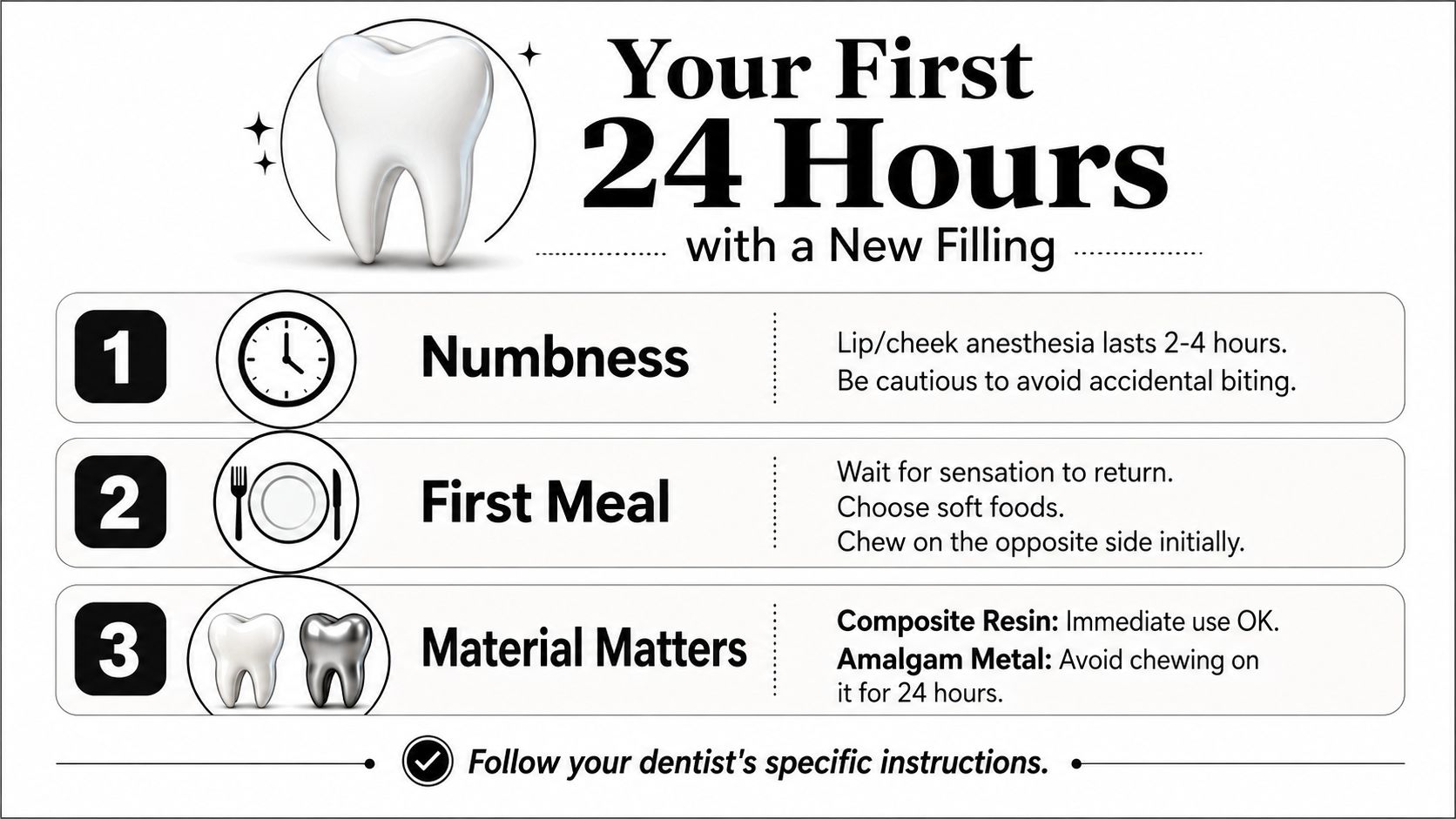
When you leave the office, the anesthetic may still affect your lip, cheek, or tongue. Don't test the numb area by chewing immediately, and be careful with hot drinks because reduced sensation can make it harder to judge temperature. Wait until normal feeling returns before eating a full meal.
Composite resin is cured in the office, so the restoration can generally be used once the numbness has resolved. Amalgam follows different handling instructions, and your dentist may recommend avoiding chewing directly on it for a settling period. Always follow the specific advice given for the material placed in your tooth.
What feels normal
Mild sensitivity to cold or pressure can occur after treatment, especially when the cavity was deep or the tooth was heavily manipulated. The tooth may feel unfamiliar because its contour and contact with the opposing tooth have changed, but that sensation should settle rather than intensify.
Brush gently along the treated area and floss as usual. When floss passes through the contact, guide it down and lift it out rather than snapping it forcefully against the gum. Avoid sticky sweets and extremely hard foods during the first day if the area feels tender, and keep sugary exposures less frequent to reduce the conditions that cause decay around restoration margins.
When to call the dentist
Spontaneous pain, throbbing, worsening sensitivity, pain that lingers, or a sharp edge deserves a call. Pain when you bite can sometimes mean the filling is high and needs a quick adjustment. Persistent symptoms may also indicate that the pulp was more inflamed than expected, so don't wait for severe pain before reporting them.
Long-term care protects both the filling and the tooth around it. Regular examinations and professional cleanings help the dentist inspect margins, contacts, and your bite. A nightguard may be useful if you grind or clench, and an early repair of a worn edge can be more conservative than waiting for a larger fracture. A dental cleaning process also gives your dental team a chance to reinforce brushing and flossing around the restoration.
Remember: A filling repairs one damaged area, but it doesn't remove the habits or conditions that caused decay. Daily fluoride care, cleaning between teeth, and attention to frequent sugary snacks help protect the tooth's remaining structure.
Paul L. Gregory, DDS offers restorative evaluations, tooth-colored composite fillings, digital diagnostics, and bite adjustments for patients who want a clear explanation of their treatment options. Visit Paul L. Gregory, DDS to schedule an assessment for sensitivity, suspected decay, or a filling that no longer feels right.