

About 39% of previously treated teeth show some sign of persistent apical disease on imaging, but most can be resolved with retreatment when the cause is identified early. A previously treated tooth may feel normal while inflammation remains around its root, so the right next step is diagnosis, not an automatic prescription, repeat procedure, or extraction.
You may be reading this after noticing tenderness when you bite, a recurring bad taste, or a small bump on your gum. Perhaps your root canal was completed months or years ago, and the tooth seemed finished until a routine X-ray raised a question. For a busy Manhattan professional, that uncertainty can be frustrating, especially when the tooth isn't causing dramatic pain.
Persistent disease after root canal treatment is commonly called post-treatment apical periodontitis. It involves inflammation and bone loss around the root, and symptoms can be mild or absent. The practical question isn't just whether an infection exists. It's whether the problem is inside the canal, outside the root, related to leakage or a fracture, and whether the tooth can be predictably saved.
When Your Root Canal Feels Wrong After Treatment
A patient we'll call Elena finished a root canal on a molar, received a crown, and returned to her normal schedule. Months later, she noticed a tender bite during lunch. The tenderness came and went, so she ignored it. Later, she detected a strange taste near the tooth and asked whether a root canal infection could return after treatment.
That pattern is common enough to deserve attention, but it doesn't automatically mean the tooth must come out. A major review of 29 prevalence studies found post-treatment apical periodontitis in 10% to 62% of patients, with a median prevalence of 39%. At the tooth level, reported prevalence ranged from 20% in a Jordanian population to 65% in a Brazilian population, with a median of 41% (international review of post-treatment apical periodontitis).
The figures don't mean that every treated tooth is actively infected or destined to fail. They show why a quiet tooth still deserves a careful examination when imaging reveals a dark area, the gum repeatedly swells, or biting remains uncomfortable. A separate review found apical periodontitis in approximately 41% of root-filled teeth, compared with 3.5% of untreated teeth (systematic review and meta-analysis).
What a symptom can and can't tell you
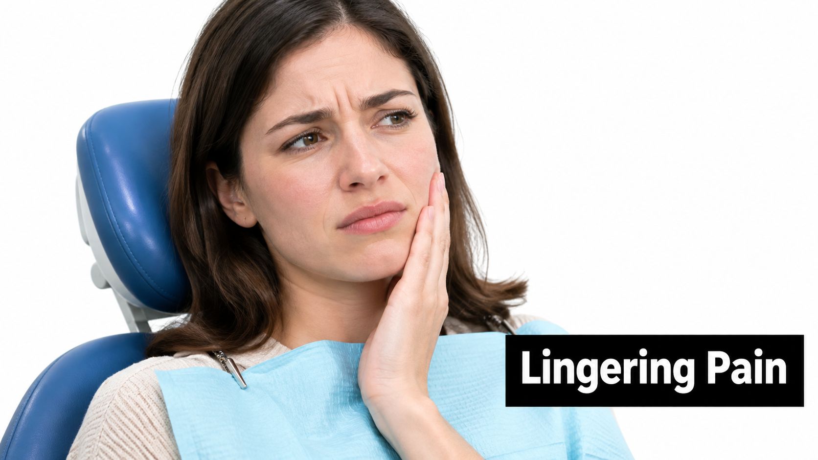
A tender bite may reflect inflammation around the root, a high crown, a crack, or pressure from surrounding tissues. A bad taste can come from drainage, gum disease, food trapping, or another nearby source. Swelling deserves prompt attention because it can indicate an abscess, particularly when it recurs.
Practical rule: Pain relief doesn't prove that the source has disappeared. A tooth that feels better may still need imaging and follow-up.
If you have facial swelling, fever, difficulty swallowing, or difficulty breathing, seek urgent dental or medical assessment. For a broader explanation of warning signs, see this guide to dental abscess symptoms and treatment.
The next step is a diagnosis that compares your current image with earlier records, checks the restoration, and evaluates the root structure. That approach helps distinguish a tooth that may benefit from nonsurgical retreatment from one better suited to apical surgery or extraction.
For a visual explanation of how persistent disease can present after treatment, review the video below.
Understanding How Infection Returns After Root Canal
A root canal removes diseased pulp, cleans the internal canal system, and seals that space. Infection can return when bacteria remain in a hidden area or regain access later. Think of the tooth as a complicated plumbing system inside a hard shell. Cleaning the visible pipe isn't enough if a side branch was missed or the outer seal breaks.
The first pathway is hidden anatomy
Molars can have narrow, curved, branching, or extra canals. A canal that wasn't found during the original procedure can retain microorganisms. Incomplete instrumentation or inadequate disinfection can leave protected areas where bacteria survive inside dentinal tubules or canal irregularities.
Persistent infections are usually polymicrobial. Reported organisms include members of the Proteobacteria, Firmicutes, Bacteroidetes, and Actinobacteria groups, with genera such as Fusobacterium, Porphyromonas, Streptococcus, and Actinomyces identified across persistent infections (review of persistent endodontic infection). The exact species matter less to a patient than the clinical implication: replacing filling material without finding the untreated space may not solve the problem.
The second pathway is leakage from above
A crown, filling, or the tooth itself can lose its seal. Saliva and oral bacteria then reach the treated chamber through a gap. Recurrent decay, a loose restoration, or restoration failure can create a new route into a previously disinfected space.
This is why the coronal seal deserves as much attention as the root filling. A well-cleaned canal can't remain protected if the opening above it permits contamination.
The third pathway is a procedural or structural problem
The original treatment may have involved limited access, insufficient cleaning, an underfilled area, or an unrecognized crack. A vertical root fracture can allow bacteria to travel along the root surface and may make conventional retreatment unreliable.
Symptoms don't always announce these problems. A lesion around the root may be visible on a radiograph before you feel pain. Your dentist may recommend imaging even when the tooth appears comfortable because clinical examination alone can't reliably exclude post-treatment disease. This overview of root canal treatment can help you understand why a completed procedure still requires a sound restoration and periodic evaluation.
The central question is therefore not, “Why did the antibiotic fail?” It's, “Where is contamination coming from, and can the source be removed?” That question guides the diagnostic workup.
Diagnostic Steps Before Deciding on Retreatment
A retreatment recommendation should follow a sequence of tests, not a guess based on one symptom or one black area on an X-ray. The clinician needs to establish whether the tooth is the source, whether the canal system can be reached, and whether the surrounding tissues suggest a different disease.
Start with the tooth, gum, and bite
The examination usually includes percussion, palpation, gum probing, mobility testing, bite assessment, and inspection of the crown or filling. A narrow, isolated periodontal pocket may raise concern about a crack. Tenderness to tapping may indicate inflammation around the root, but it doesn't identify the cause by itself.
Your dentist may also compare the treated tooth with neighboring teeth. Pain can be referred, and a nearby tooth may be responsible. The history matters too. Tell the clinician when the symptoms began, whether they appear during chewing, whether swelling drains, and whether the restoration has chipped or loosened.
Use two-dimensional images first
Periapical radiographs can reveal bone changes around the root, the length and density of the existing filling, and obvious untreated anatomy. Earlier images are valuable because a stable scar, a shrinking lesion, and a newly developing lesion can look different over time.
Digital imaging supports efficient comparison and may reduce the need for repeated conventional film. This explanation of digital X-rays for teeth describes why image quality and clinical context matter more than viewing a single photograph in isolation.
Add CBCT when the decision is uncertain
Cone-beam computed tomography, or CBCT, creates a three-dimensional view of the tooth and surrounding bone. It may reveal an extra canal, a missed root, a perforation, a fracture pattern, or a bone defect that a two-dimensional image doesn't show clearly.
CBCT isn't automatically necessary for every follow-up. The clinician weighs the expected diagnostic value against radiation exposure and uses it when the result could change the treatment plan. Imaging also has limits. It may suggest a problem, but it can't always prove that an external biofilm is causing the lesion.
Decide where the infection sits
If the suspected source is inside a reachable canal, nonsurgical retreatment may make sense. If the canal is adequately treated but disease remains at the root tip, apical surgery may offer direct access to the affected tissue. If a vertical fracture or severe structural loss prevents a durable restoration, extraction may be more predictable.
That distinction matters because a failed root canal doesn't always mean infection remains inside the canal. Extraradicular biofilms can exist on the external root surface, in apical ramifications, or within a lesion. Confirming that source may require surgical access and examination of the root tip and associated tissue (review of refractory apical periodontitis).
Choosing Between Retreatment, Surgery, or Extraction
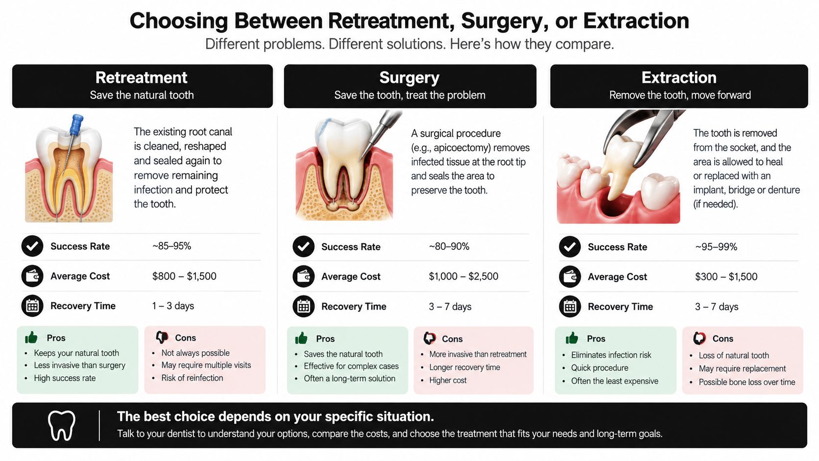
Once the examination and imaging are complete, the decision usually comes down to three paths. The best option depends on whether the tooth is structurally sound, whether the canal can be disinfected, and whether the cause of failure can be corrected.
| Option | What happens | When it tends to fit |
|---|---|---|
| Nonsurgical retreatment | Existing filling material is removed, canals are located and cleaned again, the space is disinfected, and a new seal is placed | The tooth is restorable and contamination appears to be inside the canal |
| Apical surgery | A small surgical access is made through the gum, the root tip and lesion are treated, and the root end is sealed | The canal is adequately treated, access from the crown is impractical, or disease may be outside the canal |
| Extraction and replacement | The tooth is removed, then replacement is planned with an implant, bridge, or denture | A fracture, severe structural damage, or poor restorability makes preservation unpredictable |
Nonsurgical retreatment
Retreatment can preserve the natural tooth while addressing missed anatomy, poor disinfection, or leakage. A contemporary review reported complete healing in approximately 74% to 86% of cases after nonsurgical retreatment. When healed teeth, healing teeth, and teeth functioning without symptoms were counted together, 91% to 97% remained in asymptomatic function (review of retreatment outcomes).
The procedure often requires removal of the existing filling material and, sometimes, removal or modification of a crown or post. The clinician then searches for untreated canals, corrects access problems, reduces microbial contamination, and creates a durable coronal seal. A tooth that can be restored properly is usually a stronger candidate than one with extensive loss of structure.
Apical surgery
Endodontic microsurgery approaches the root through the gum rather than reopening the entire canal from above. The surgeon can remove the root tip and associated inflamed tissue, inspect the area under magnification, and place a root-end seal.
This option becomes more relevant when a canal is blocked, a post makes crown access risky, or the canal appears adequately treated while a lesion persists. The cited review reports average four-year success rates of approximately 77% to 91% for endodontic microsurgery (review of extraradicular infection and microsurgery). It isn't a punishment for a failed root canal. In selected cases, it provides direct access to a problem that instruments cannot reach from inside the tooth.
Extraction
Extraction removes the tooth and its internal and external sources of infection. It may be the soundest choice when a vertical root fracture, inadequate remaining tooth structure, severe periodontal support loss, or an unmanageable restoration makes long-term sealing unlikely.
Replacement planning still matters. An implant, bridge, or denture has its own surgical, restorative, and maintenance considerations. Ask what happens to the neighboring teeth, the bone, your bite, and your schedule before choosing removal only because it appears faster.
A reasonable consultation should explain not only what can be done, but why one route is more predictable for your tooth.
If you want to compare preservation and replacement in plain language, read about root canal versus extraction. Paul L. Gregory, DDS offers evaluation, digital diagnostics, root canal treatment, and retreatment when indicated, with restorative and implant planning available when a tooth can't be predictably preserved.
Pain Management and Recovery Timeline After Retreatment
Most patients worry about two separate experiences: discomfort during the procedure and soreness afterward. Local anesthesia is used during treatment, and the clinician can discuss anxiety-control options when fear makes dental care difficult. Afterward, tenderness can occur because the tissues around the root have been irritated and are beginning to settle.
The first day
Follow the instructions from your treating dentist rather than combining medications on your own. Over-the-counter anti-inflammatory medicine may be appropriate for some adults, but allergies, pregnancy, stomach conditions, kidney disease, blood thinners, and other medicines can change what is safe.
Eat carefully until normal sensation returns. Avoid chewing hard foods on the treated tooth until your dentist confirms that the temporary or final restoration can tolerate normal function. Mild soreness with biting can occur during early healing.
The next few days
Many people return to ordinary activities within one to two days, but individual recovery varies. Swelling that increases, severe pain that doesn't respond to the recommended plan, fever, a spreading facial swelling, or difficulty swallowing requires prompt contact with the dental office or urgent assessment.
A short course of antibiotics isn't a substitute for source control. CDC guidance summarizing ADA recommendations states that antibiotics are generally unnecessary for most dental pain and intraoral swelling in immunocompetent adults when definitive dental care is available. They become more relevant when systemic involvement, such as fever, malaise, or spreading cellulitis, is present.
The healing period
Pain often settles before the bone around the root has fully repaired. Radiographic healing can take three to six months, so follow-up imaging may be scheduled later rather than immediately. A gradual reduction in tenderness, no recurring swelling, and comfortable chewing are reassuring clinical signs, but the dentist uses both symptoms and imaging to assess progress.
For a practical recovery checklist, see this guide to root canal recovery. If symptoms improve after antibiotics but return, don't assume the infection has been eradicated. Arrange reassessment so the underlying canal, restoration, fracture risk, or apical lesion can be addressed.
Prognosis Factors and When to Seek a Second Opinion
The strongest predictor isn't the word “retreatment” on the treatment plan. It's whether the clinician can identify and correct the reason the first procedure failed. A tooth with missed anatomy and a sound outer structure presents a different problem from a tooth with a vertical fracture or a restoration that can't be sealed.
Important questions include:
- Cause: Is the problem missed anatomy, inadequate cleaning, leakage, new decay, a fracture, or disease outside the root?
- Restorability: Can enough healthy tooth remain to support a reliable crown or other final restoration?
- Anatomy: Can the canals be reached safely, or would surgery provide more direct access?
- Sealing: Will the new restoration prevent saliva and bacteria from re-entering?
- Surrounding tissues: Does the lesion or periodontal condition alter the likely outcome?
Historical studies of retreatment report pooled success rates of approximately 76.6% to 78%, with published results ranging from 62% to 86%, and one report found 89% five-year survival for retreatment cases (American Association of Endodontists review). These figures describe groups, not a personal guarantee. Your diagnosis, anatomy, restoration, and treatment quality determine where your case fits.
A second opinion is sensible when extraction is recommended without a clear explanation of the fracture or restorability, when no one has reviewed prior radiographs, or when retreatment and apical surgery haven't been compared. It can also help when symptoms persist despite apparently adequate canal treatment, because an external lesion may require a different approach.
Manhattan patients may use neighborhood names such as Midtown, Chelsea, the Upper West Side, the Upper East Side, Lenox Hill, Turtle Bay, Hell's Kitchen, the Theatre District, Gramercy Park, West Village, Greenwich Village, NoHo, East Village, SoHo, Tribeca, Hudson Yards, Chinatown, and Wall Street. These locations can help you find convenient care, but they don't define identical official boundaries. New York City uses 59 community districts, and Tribeca, the Financial District, and Battery Park City are within Community Board 1 (New York City Department of City Planning community-district information).
If you have lingering tenderness, swelling, drainage, or an X-ray finding after root canal treatment, schedule a diagnosis-focused visit rather than waiting for pain to become severe. Paul L. Gregory, DDS can evaluate a suspected root canal retreatment infection, use digital imaging to assess the tooth, and coordinate retreatment, restoration, or replacement planning when appropriate. Visit Paul L. Gregory, DDS to request an evaluation in Midtown Manhattan.