

A 19-year-old notices tightness behind the lower molar while chewing. The next morning, the gum behind that tooth is swollen, tender, and leaving a metallic taste. The immediate question is simple: when should wisdom teeth be removed?
In practice, the answer rarely comes from age alone. I assess the tooth's angle, the amount of room available, the condition of the neighboring second molar, the presence of gum inflammation or decay, and what the X-ray shows. A teenager with a severely tilted tooth may benefit from removal before symptoms worsen, while an older patient with a healthy, fully erupted, easy-to-clean tooth may have no reason to undergo surgery.
Wisdom teeth, also called third molars, usually erupt between 18 and 24. A review referenced by the National Institute for Health and Care Excellence notes that about 80 out of 100 young adults have at least one wisdom tooth that remains inside the jaw. That makes the decision common, but it doesn't make every case identical.
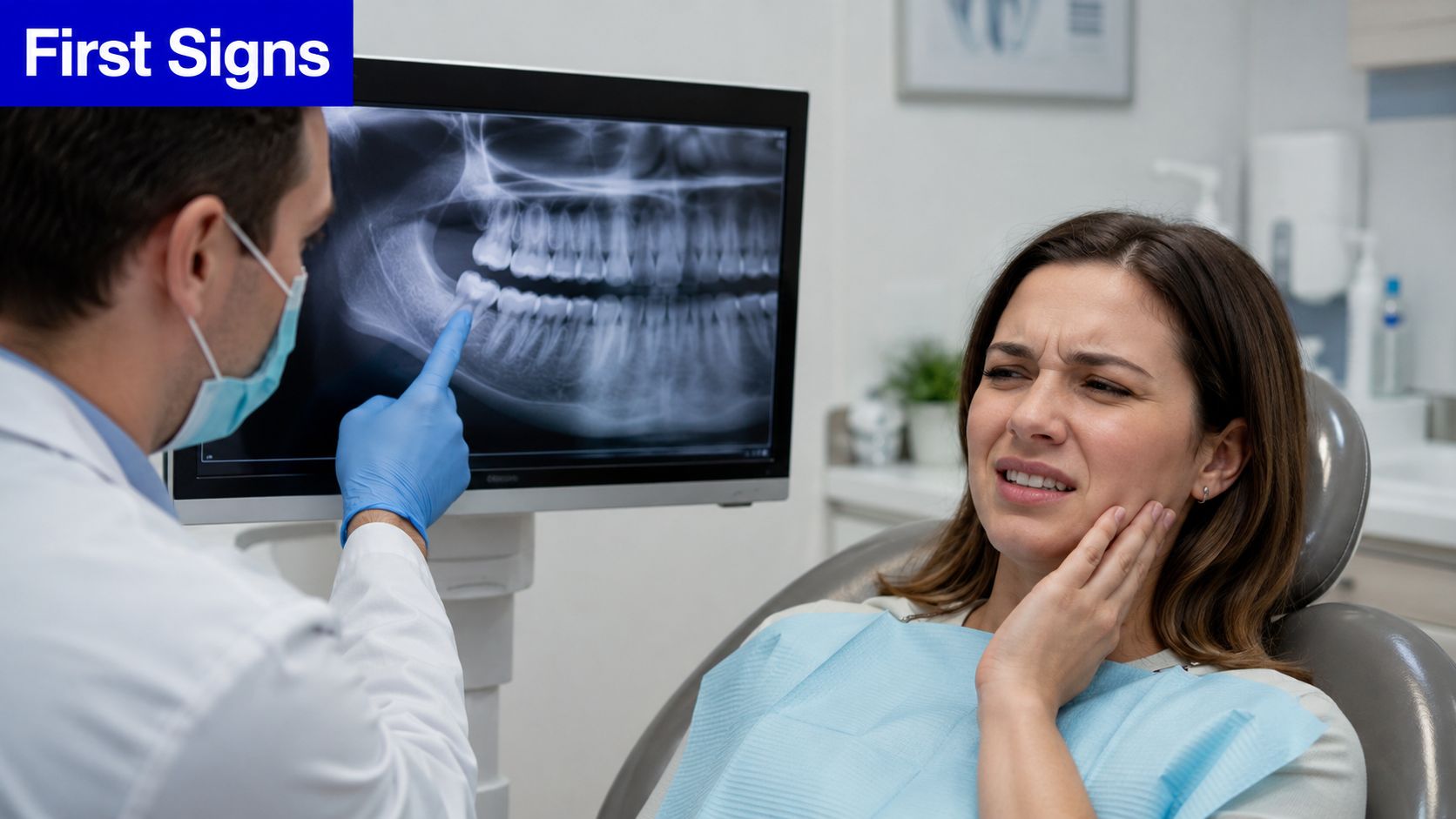
If your child or teenager is approaching the age when third molars become visible on imaging, a pediatric dental evaluation can establish whether the teeth are developing normally. The useful question isn't whether the teeth are present. It's whether they're likely to remain healthy, accessible, and harmless.
The Moment Most People First Wonder
The first episode often feels minor. You may notice pressure while biting, a dull ache at the back of the jaw, or a flap of gum that catches food. Then the area becomes difficult to brush, the jaw feels stiff, and swelling appears around the last molar.
That pattern can indicate pericoronitis, inflammation around a partially erupted wisdom tooth. The tooth may have enough space to show part of its crown but not enough room to emerge fully. Food and bacteria collect beneath the gum, and the area becomes a recurring source of irritation.
The decision is rarely binary
Patients often ask whether every wisdom tooth should come out. A safer approach is to classify the tooth according to risk:
- Low risk: The tooth is fully erupted, functional, cleanable, and shows no decay or gum damage.
- Intermediate risk: The tooth is partially erupted or difficult to clean, but current disease is limited.
- High risk: The tooth is severely angled, damaging the second molar, associated with recurrent inflammation, or surrounded by radiographic pathology.
The American Dental Association's patient guidance describes removal as a response to pain, gum disease, or other evidence of change in the mouth, rather than age alone. That distinction matters. A painless tooth can still show measurable damage, while a brief episode of soreness doesn't automatically make extraction necessary.
Practical rule: Pain gets your attention, but the X-ray and periodontal examination usually determine the timing.
I also consider age because surgical difficulty tends to increase as patients get older. A major clinical review associated with UK guidance reports that 70% to 80% of mandibular third molars had been removed by age 38, and that more than 80% required removal before age 38. Those figures show that many teeth are removed before middle age, but they don't prove that every tooth should be extracted immediately.
What Impaction Actually Looks Like
An impacted wisdom tooth is blocked from reaching a normal position in the mouth. The blockage may come from the second molar, the jawbone, the gum, or a lack of space.
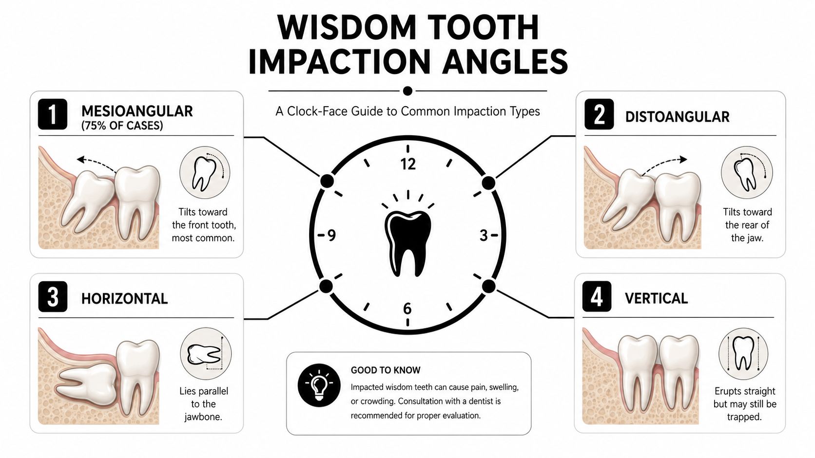
On a panoramic X-ray, imagine the second molar standing upright like the number 12 on a clock. The wisdom tooth's crown then points in a particular direction. That direction helps predict what structures may be at risk and how difficult removal could become.
Four positions clinicians commonly describe
- Mesioangular: The crown tilts forward toward the second molar. Reviews commonly describe this as the most frequent impaction pattern. The contact point can trap plaque against the second molar's back surface, creating distal cervical decay or periodontal damage.
- Distoangular: The crown leans backward toward the ramus. The tooth may sit deeper in bone and can be technically more demanding to remove because the surgeon has less direct access.
- Horizontal: The tooth lies on its side, with the crown directed into the second molar's root or crown. This arrangement can place pressure on the neighboring tooth and may be associated with root resorption or cystic change.
- Vertical: The tooth is upright but remains trapped because the jaw lacks sufficient eruption space. A vertical tooth isn't automatically harmless. Its depth, gum coverage, and relationship with the second molar still matter.
The angle is only one part of the assessment. A mesioangular tooth with a shallow crown and obvious contact against the second molar may deserve earlier attention than a deeper vertical tooth with a stable appearance. Conversely, a horizontal tooth with no current symptoms may still require removal if the image shows damage developing around it.
The NIH review of wisdom tooth management identifies high-risk patterns such as horizontal or severely mesioangular impaction, recurrent pericoronitis, caries, distal periodontal injury, cystic change, and other local disease. These findings convert an abstract concern into a clinical indication.
Symptoms Worth Acting On
A single twinge doesn't tell me much. A repeatable symptom pattern, especially when it corresponds to a visible abnormality, carries far more weight.
Symptoms that change the conversation
Pericoronitis causes tenderness, swelling, and irritation around a partially erupted tooth. A bad taste may come from trapped debris or drainage from inflamed tissue. When inflammation keeps returning despite careful cleaning, the tooth's anatomy may be the problem rather than the patient's effort.
Sensitivity behind the second molar deserves close attention. A mesioangular wisdom tooth can press against the neighboring molar in a way that makes the back surface difficult to inspect and clean. By the time a patient feels cold sensitivity or localized pain, decay may already be established.
Intermittent swelling can signal a pocket around the tooth, recurrent infection, or less commonly a cystic process. A panoramic image helps identify whether the follicular space around the crown looks unusual or whether the tooth is affecting nearby roots and bone.
Bleeding from the gum flap is another useful clue. It suggests inflammation in an area that may be difficult to keep clean, particularly when the tooth is only partly erupted.
Match the symptom to the next action
- One mild episode: Arrange a routine examination and imaging if the tooth hasn't been assessed recently. The immediate goal is to determine whether the problem is local and reversible.
- Repeated inflammation: Discuss removal with a dentist or oral surgeon, especially if the same tooth repeatedly traps food or causes swelling.
- Second-molar sensitivity or visible decay: Request an evaluation of both teeth. Removing the wisdom tooth alone may not resolve damage that already affects the second molar.
- Swelling with restricted opening, pus, fever, or swallowing difficulty: Seek urgent dental assessment. These findings can indicate an infection that needs prompt treatment.
Patients with sudden jaw swelling or significant pain can use an emergency dental assessment rather than waiting for a routine cleaning. Antibiotics may sometimes be part of initial care, but they don't eliminate an impacted tooth that continues to create a bacterial trap.
How Imaging Shapes the Decision
The first useful image is often a panoramic radiograph. It shows both jaws and gives a broad view of the wisdom teeth, second molars, surrounding bone, and the course of important anatomical structures.
I use that image to assess the wisdom tooth's angulation, depth, relationship with the second molar, crown-to-bone position, gross root shape, and any obvious radiolucency. It can also reveal whether the second molar has distal caries or whether the wisdom tooth is pressing against its root.
Each view answers a different question
A periapical film provides a more focused image of a particular tooth and its surrounding bone. It can help clarify subtle distal bone loss, early root resorption, or the cervical area of the second molar when a panoramic view isn't sufficiently detailed.
A CBCT scan creates a three-dimensional view. I don't order it automatically for every wisdom tooth. It becomes useful when a panoramic image suggests close proximity to the inferior alveolar nerve, when roots appear curved or dilacerated, or when unexplained pathology requires a more precise map before surgery.
| Modality | Best for | What it reveals | When to order |
|---|---|---|---|
| Panoramic radiograph | Initial third-molar screening | Angulation, depth, neighboring tooth relationship, gross roots, visible pathology | Baseline assessment or a change in symptoms |
| Periapical radiograph | Focused evaluation | Distal bone, cervical decay, root resorption, localized detail | When the panoramic image leaves a specific concern unresolved |
| CBCT | Three-dimensional surgical planning | Nerve proximity, root curvature, cortical boundaries, pathology extent | When two-dimensional imaging suggests elevated surgical risk or unclear disease |
The purpose of imaging isn't to collect pictures. It's to decide whether removal is justified now, whether a specialist should plan the procedure, or whether a stable tooth can enter surveillance.
Patients who want to understand how modern dental imaging works can review information about digital X-rays for teeth. Your clinician should be able to point to the exact radiographic feature driving the recommendation.
Why Age 17 to 25 Keeps Coming Up
The late teens through early twenties often provide the most favorable balance between tooth development, bone conditions, and surgical planning. That doesn't mean every wisdom tooth should be removed during this period. It means that when extraction is indicated, delaying without a clear surveillance plan may reduce the margin for an uncomplicated procedure.
Wisdom teeth usually erupt between 18 and 24, according to the NICE clinical-need review. The same review explains why late-teen and young-adult timing has historically been favored, extraction is often easier before the teeth fully develop and the roots lengthen.
The age curve needs an honest explanation
A PubMed-indexed review reports that older patients tend to have longer postoperative courses and more complications. Another study associates age above 25 with increased complication risk, with an odds ratio of 1.5, as summarized in the oral surgery review indexed by Wiley.
That doesn't make age 25 a rigid deadline. It's better understood as a practical inflection point. As roots lengthen and may curve, the tooth can become more firmly positioned in bone. The surrounding bone and the nerve relationship also influence the difficulty of removal.
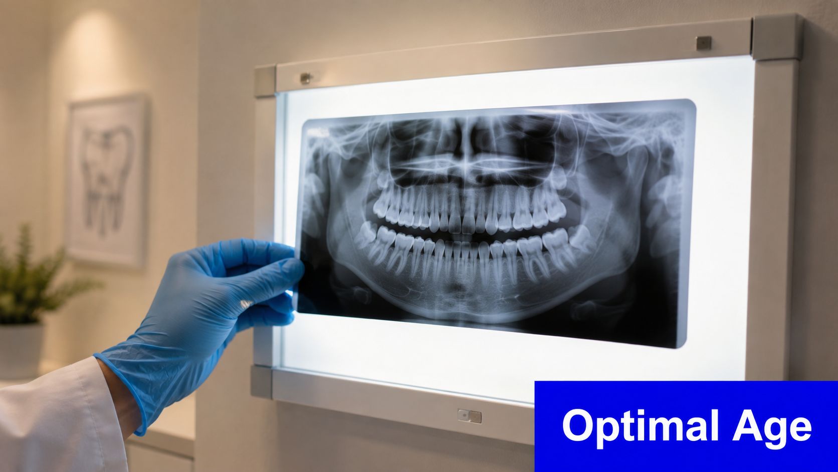
For an adolescent whose X-ray shows a horizontal or severely mesioangular tooth, earlier surgery may prevent damage to the second molar and avoid a more difficult extraction later. For a healthy, fully erupted tooth, surgery may offer little benefit and exposes the patient to operative risk without a clear disease target.
Orthodontic planning can also affect timing. Families considering braces for teens should ask whether the third molars are relevant to the treatment plan, but they shouldn't assume that orthodontic treatment alone proves extraction is necessary.
Removing Versus Keeping Wisdom Teeth
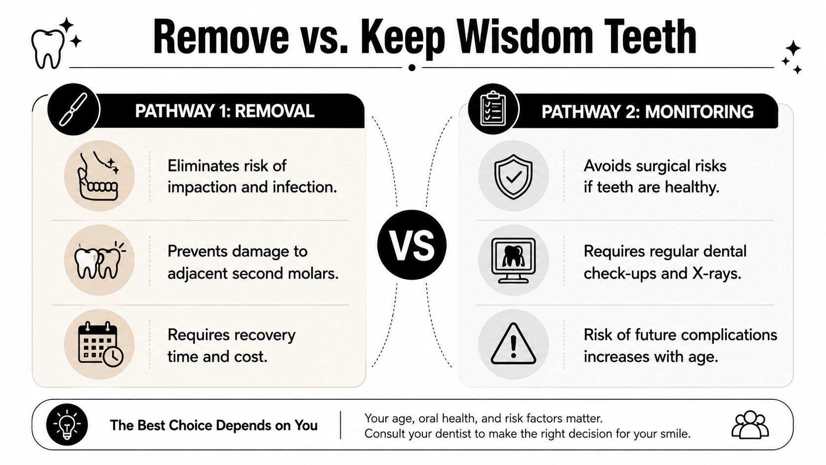
There are two legitimate pathways, removal and monitored retention. The correct choice depends on whether the tooth is healthy and maintainable, not on a universal rule that applies to every patient.
What removal solves
Extraction can eliminate a source of recurrent pericoronitis, remove an impacted tooth that is damaging the second molar, and address a cystic or other pathological change. It may also be more predictable when performed earlier in young adulthood, before the roots lengthen and the surgical relationship becomes less favorable.
The trade-off is immediate. Surgery carries risks such as postoperative pain, swelling, infection, and possible nerve irritation. The risk profile depends on tooth position, root anatomy, nerve proximity, medical history, and the clinician's surgical plan.
What retention preserves
Keeping a wisdom tooth avoids surgical morbidity when the tooth is healthy, erupted, functional, and easy to clean. It also respects a patient's preference when clinical and radiographic findings show no meaningful disease risk.
Retention only works when the tooth can be examined and maintained. A fully erupted tooth that participates in chewing and allows effective brushing is different from a heavily impacted tooth that cannot be inspected directly.
The absence of pain doesn't equal the absence of pathology.
The asymptomatic but pathological middle ground
Many simplified explanations fail here. A patient may feel completely comfortable while an X-ray shows radiolucency around an impacted crown, early root resorption on the second molar, or progressive distal bone loss.
The NICE guidance on wisdom tooth extraction recommends discontinuing prophylactic removal of pathology-free impacted third molars. A later evidence review notes that the American Association of Oral and Maxillofacial Surgeons supports surgical management when erupted or impacted teeth have present or potential pathology, even without symptoms.
Those positions appear conflicting until the word pathology is made explicit. The question isn't “Does it hurt?” It's “Does the tooth show caries, periodontal disease, cystic change, resorption, distal bone loss, or interference with other treatment?”
What Watchful Waiting Really Means
Watchful waiting isn't a decision to forget about the tooth. It's active surveillance, with a written baseline and planned reassessment.
A 2024 clinical guideline review recommends clinical checks every 6 to 12 months for retained third molars, with panoramic radiographs about every 2 years when abstention is chosen, as described in the NIH guideline review. The exact interval may change if the tooth is partly erupted, difficult to clean, or already showing a suspicious finding.
What the clinician checks
At each clinical visit, the dentist should assess the gum around the second and third molars, look for food trapping, inspect the second molar's distal surface, and compare symptoms with the previous record. Periodontal probing helps identify whether the pocket behind the second molar is becoming deeper or harder to maintain.
Radiographs provide a different layer of information. The clinician compares the tooth's position, surrounding bone, follicular space, adjacent-root relationship, and evidence of decay or resorption over time.
When a retained tooth develops a documented indication, surveillance should end and the treatment plan should change. That may mean extraction, additional imaging, referral to oral surgery, or treatment of the second molar if damage has already occurred.
Monitoring requires patient participation
A surveillance plan fails if appointments are missed or if new symptoms are ignored. Patients should report swelling, bad taste, bleeding, difficulty opening the mouth, or new sensitivity behind the second molar rather than waiting for the next scheduled review.
For patients who have difficulty attending in person, a teledentistry consultation may help with preliminary discussion and record review. It can't replace the clinical examination or required imaging, but it can help clarify whether an in-person appointment needs to be routine or urgent.
Your Next Step Based on Where You Are Now
Use three facts to guide the next appointment: your age, your symptoms, and the date and quality of your latest imaging. Those variables produce a more useful plan than a blanket recommendation to remove every wisdom tooth.
A practical decision tree
- Age 16 to 25, no symptoms: Arrange a baseline panoramic assessment if one hasn't been completed recently. If the teeth appear healthy but their future position is uncertain, establish active surveillance rather than scheduling surgery automatically.
- Any age with symptoms: Request a clinical assessment and appropriate imaging promptly. A panoramic image may be paired with a focused periapical view when the second molar or distal bone is a concern, and an oral-surgery referral may be appropriate when impaction or pathology is present.
- Age 35 or older, stable and asymptomatic on recent imaging: Continue clinical review and compare future images for change. Retention can remain reasonable when the tooth is stable, cleanable, and free of disease, but age alone shouldn't substitute for examination.
- Facial swelling, pus, restricted mouth opening, or sudden numbness: Seek same-day dental or maxillofacial assessment. These findings can indicate infection or involvement of important nearby structures and shouldn't wait for a routine cleaning.
The decision to see an oral surgeon becomes stronger when the tooth is horizontal or severely mesioangular, the second molar shows distal damage, the roots appear close to the inferior alveolar nerve, or a cystic change is suspected. A general dentist can often monitor a healthy, accessible tooth and coordinate referral if the findings change.
The most useful question to ask at your appointment is: “What specific finding makes removal safer than monitoring in my case?” If the answer points to a visible risk marker, timing matters. If the answer is that the tooth is healthy and pathology-free, ask for the surveillance interval and the exact changes that would trigger removal.
Paul L. Gregory, DDS offers examinations, digital imaging, routine and surgical extractions, and wisdom teeth removal consultations in Manhattan, including care planning for patients who are anxious about treatment. Visit Paul L. Gregory, DDS to arrange an evaluation based on your symptoms, age, and latest X-ray findings.